X-Rays
A Diagnostic Tool for Comprehensive Oral Health Assessment
Dental X-rays, also known as radiographs, play a crucial role in dentistry as an essential diagnostic tool. They provide detailed images of the teeth, jawbone, and surrounding tissues, enabling dentists to assess oral health comprehensively. Here’s a comprehensive overview of dental X-rays:
1. Types of Dental X-Rays:
- Bitewing X-Rays: Capture images of the upper and lower teeth in a specific area, showing details of the crowns and supporting bone.
- Periapical X-Rays: Focus on one or two specific teeth, providing a detailed view of the entire tooth from crown to root and the surrounding bone.
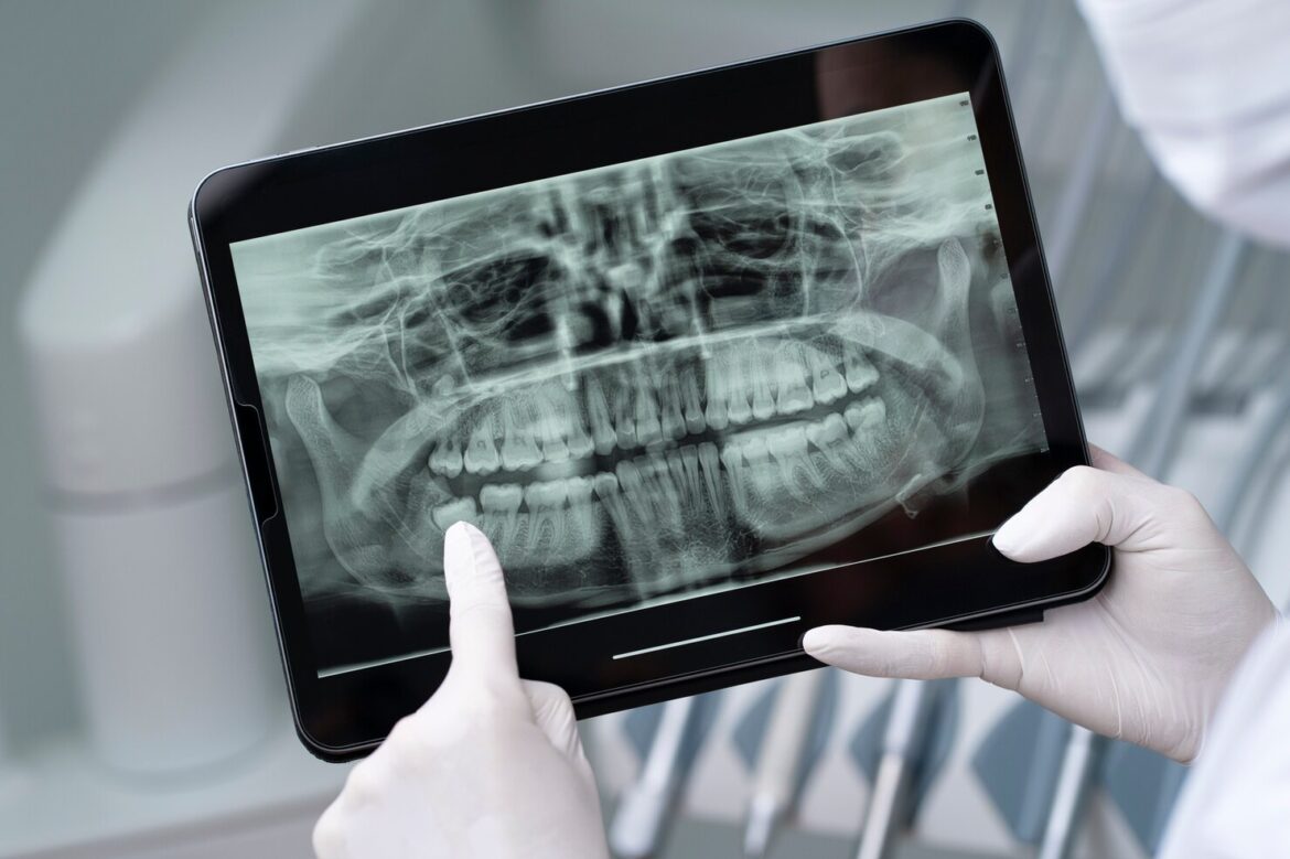
- Panoramic X-Rays: Capture a broad view of the entire mouth, including all teeth, jaws, and surrounding structures.
- Orthodontic X-Rays: Used to assess the alignment of teeth and jaws for orthodontic treatment planning.
- Cone Beam Computed Tomography (CBCT): A three-dimensional imaging technique providing detailed views of oral and facial structures.
2. Purpose and Importance:
- Diagnosis of Dental Issues: X-rays help identify cavities, infections, cysts, tumors, and other dental abnormalities that may not be visible during a clinical examination.
- Evaluation of Tooth Roots: X-rays reveal the structure and health of tooth roots, aiding in the diagnosis of root canal issues.
- Assessment of Bone Density: X-rays are essential for evaluating the density and health of the jawbone, crucial for procedures like dental implant placement.
- Orthodontic Treatment Planning: X-rays assist orthodontists in planning and monitoring the progress of orthodontic treatments.
3. Frequency of Dental X-Rays:
- Individualized Approach: The frequency of X-rays depends on individual oral health needs, age, and risk factors.
- New Patients: X-rays are often taken for new patients to establish a baseline and identify any pre-existing conditions.
4. Safety Measures:
- Low Radiation Exposure: Modern dental X-ray equipment minimizes radiation exposure, and lead aprons and thyroid collars are used to protect patients from unnecessary radiation.
- Pregnancy Precautions: Pregnant individuals may avoid routine X-rays, and if necessary, appropriate shielding is employed.
5. Advancements in Technology:
- Digital X-Rays: Digital imaging reduces radiation exposure and allows for instant image processing.
- Cone Beam CT (CBCT): Provides detailed three-dimensional images, particularly useful for complex dental procedures.
6. Patient Involvement:
- Informed Consent: Dentists typically obtain informed consent before performing X-rays, explaining the purpose and benefits to the patient.
7. Environmental Impact:
- Digital Transition: The shift to digital X-rays has reduced the environmental impact associated with traditional film processing.
Dental X-rays are an invaluable tool in modern dentistry, enabling dentists to diagnose and treat a wide range of oral health issues effectively. The benefits of early detection and precise assessment provided by X-rays contribute significantly to maintaining and improving overall oral health. If you have concerns or questions about dental X-rays, discuss them with your dentist to ensure a well-informed and personalized approach to your oral care.
